Fig. 4.
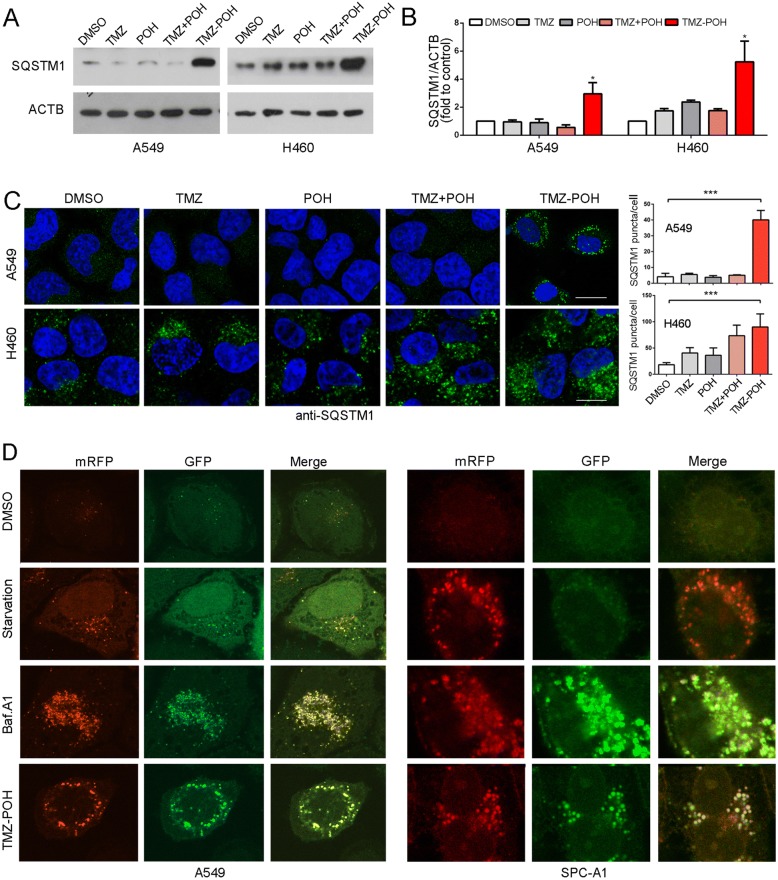
TMZ-POH blocks mitophagy flux. a-c Cells were treated with 100 μM TMZ, POH, TMZ + POH, TMZ-POH or DMSO respectively for 48 h in A549 and H460 cells. (A-B) Western blot analysis demonstrated SQSTM1 expression; (c) The above drug-treated cells were inspected under confocal laser microscopy to detect SQSTM1 puncta by immunofluorescence and SQSTM1 puncta number per cell was quantified using the Fiji Image J program. d A549 and SPC-A1 cells expressing mRFP-GFP-LC3 were starved or treated with Baf.A1 or TMZ-POH, and imaged by confocal microscopy. The results shown are means ±SD, *p < 0.05, **p < 0.005, ***p < 0.001

