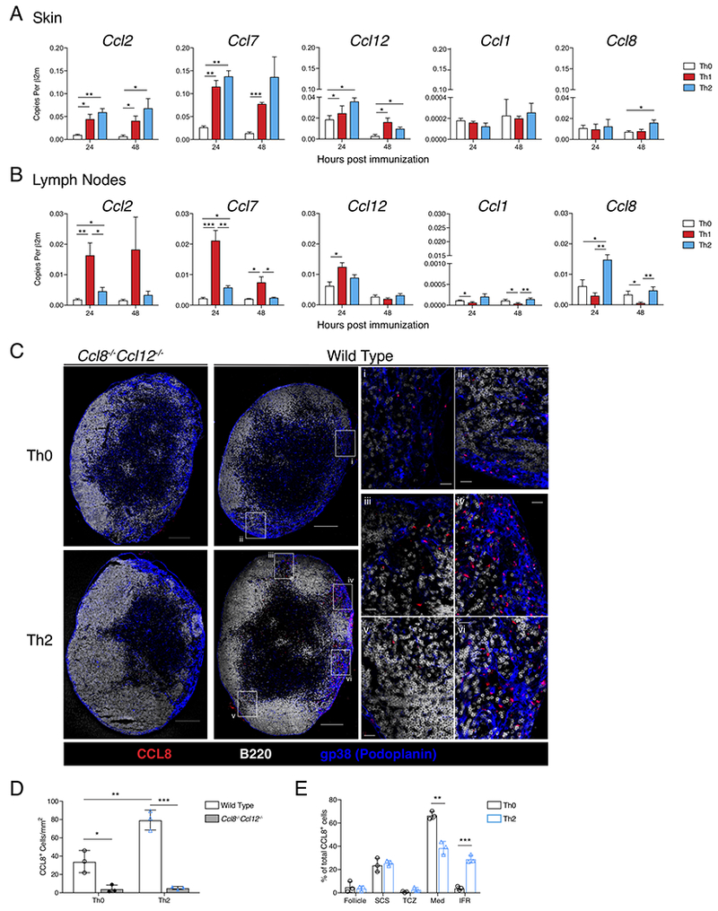
Figure 2: Allergen immunization promotes a specific chemokine signature in the dLN.

(A) QPCR analysis of footpad skin immunization site and (B) popliteal LN, 24 and 48 hrs after immunization with Th0-, Th1-, or Th2-skewing immunizations. Data is expressed as cDNA copies of indicated gene per copies of β2m. Each bar represents the mean of 3-6 mice from a representative of 4 independent experiments. Error bars indicate SEM. (C) CCL8 protein staining in whole LNs. Popliteal LNs of WT or Ccl8−/−Ccl12−/− mice were harvested 24 hrs after immunization. Confocal immunofluorescence of whole LNs shows CCL8 (red), B220 (white), and gp38 (blue), scale bar indicates 200μm. Insets show medullary (i) and medullary/follicular border (ii) in Th0-immunized WT mouse. In Th2-immunized WT mouse, insets show interfollicular regions (IFR) (iii, iv, and v) and medullary region (vi). Scale bar in insets indicates 20 μm. (D) Total CCL8+ cells per mm2 of LN section from WT and Ccl8−/−Ccl12−/− mice 24 hours after Th0 or Th2-immunization. (E) Percent of CCL8+ cells in each indicated region out of total CCL8+ cells per LN section in wild type Th0 or Th2-immunized mice: Follicle (B cell follicle), SCS (subcapsular sinus), TCZ (T cell zone), Med (medullary region), IFR. Data shown is a representative section from 3 mice used in each of 3 independent experiments. * p<0.05, ** p<0.01, *** p<0.001, Student’s T test. See also Figure S2.
