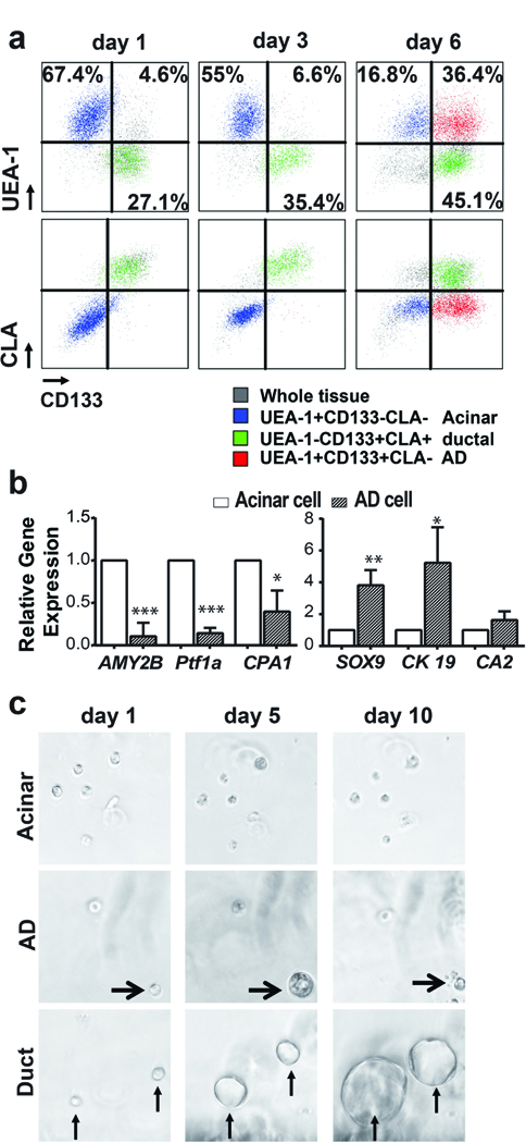
FIGURE 1. Characterization of ADM of primary human pancreatic acinar cells in vitro.

a) Cells of acinar origin (blue, UEAhighCD133−) gained CD133 expression during culture to become CD133+ cells (red, UEAhighCD133+), but continued to be CLA negative. Cells of ductal origin (green, UEAlowCD133+) showed no significant change in CD133 and CLA expression during culture (n=8). b) AD cells expressed significantly higher levels of ductal-specific genes (CK19 and SOX9) and lower levels of acinar-specific genes (AMY2B and PTF1a) than acinar cells (n=3). Data represents mean (SD) from three independent experiments. *P < 0.05, **P < 0.01, ***P < 0.001. c) Sphere formation assay for cells sorted from 2D culture. Ductal cells proliferated continuously to form large spheres and acinar cells could not form spheres, AD cells acquired the ability to proliferate transiently to form small spheres (arrowhead).
