
FIGURE 3.
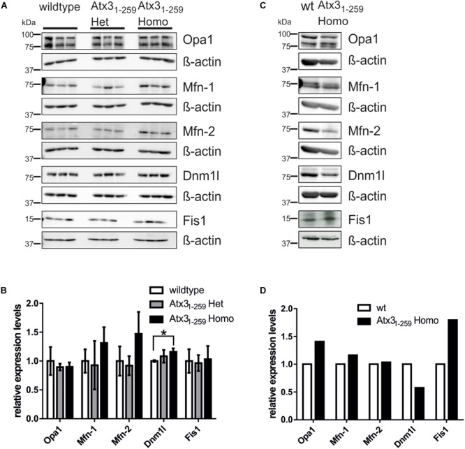
Abnormalities in mitochondrial morphology are not mainly caused by differential expression of fission and fusion proteins. (A,C) Western blot analyses in whole brain lysates of 12 months old mice (3 animals per genotype; A,B) or MEF lysates (one sample per genotype; C,D) represent an equal expression of fission (Dnm1l and Fis1) and fusion proteins (Mfn-1, Mfn-2 and Opa1). β-actin is shown as loading control. (B,D) Densitometric quantification revealed significantly increased Dnm1l protein levels in mice homozygous for truncated Atx31-259 compared to wildtype (p = 0.028).