
FIGURE 7.
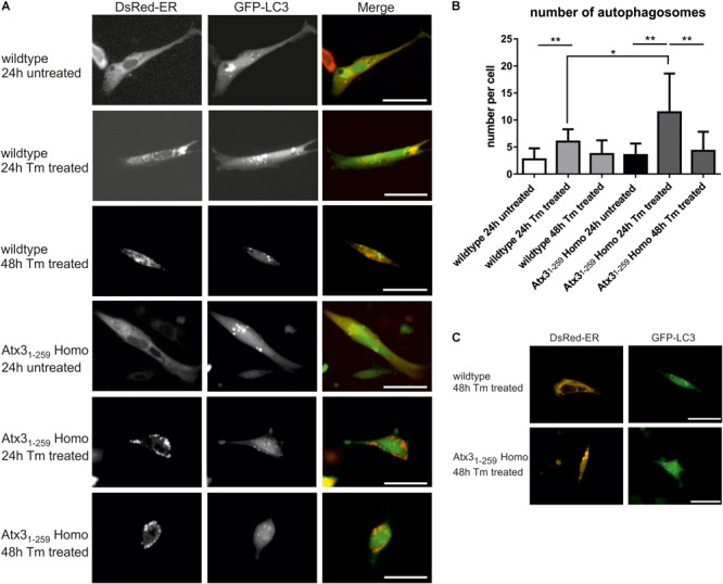
Mild ER stress leads to early autophagy activation but still results in accumulation of unfolded proteins within the ER in homozygous Atx31-259 cells. (A) Living wildtype and homozygous Atx31-259 MEF were transfected with pDsRed-ER and pEGFP-LC3 vectors and treated with 1 μg/ml tunicamycin (Tm) for 24 and 48 h. Wildtype cells reacted with producing more autophagosomes after 24 h of treatment and came back to normal steady-state autophagy levels after 48 h of treatment. The ER was not affected. In comparison, homozygous Atx31-259 MEF also started with producing more autophagosomes after 24 h of treatment but at the same time point unfolded proteins started to accumulate within the ER. This accumulation resulted in “bubble-like” DsRed-positive structures found in homozygous Atx31-259 MEF. After 48 h of treatment the accumulation of ER proteins was constantly going on but the number of autophagosomes resembled the number of autophagosomes detected under untreated conditions. Bar indicates 20 μm, represented pictures of three independent experiments is shown. (B) Counting the number of autophagosomes in 50 cells from three independent experiments revealed a significantly higher number of autophagosomes in 24 h Tm-treated wildtype and homozygous Atx31-259 cells compared to untreated cells and cells, which were treated for 48 h with tunicamycin. Additionally, the number of autophagosomes was significantly higher in homozygous Atx31-259 MEF compared to wildtype cells after 24 h of treatment (∗p < 0.05, ∗∗p < 0.01). (C) As a control experiment, cells were transfected with either pDsRed-ER or pEGFP-LC3 and treated with 1 μg/ml tunicamycin for 48 h. Comparable results were seen as demonstrated for double-transfected wildtype and homozygous Atx31-259 cells. Scale bar indicates 20 μm.