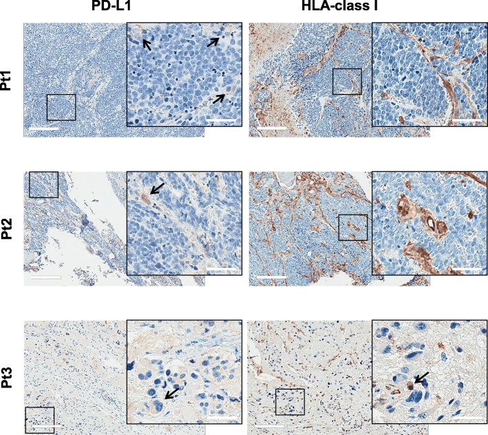
Fig. 4.
PD-L1 and HLA-class I expression in NBL before the vaccination. IHC was performed on consecutive sections of FFPE tumor samples before the vaccination. Representative IHC images of PD-L1 and HLA-class I markers for the three enrolled patients (Pt1, 2, 3) are reported. Arrows indicate PD-L1 and HLA-class I positive tumor cells. Scale bar = 200 μm; inserted panels scale bar = 50 μm

