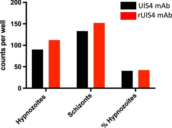
Fig. 3.

Comparison of liver stage schizont and hypnozoite counts using α-rUIS4 mAb or α-UIS4 hybridoma-derived mAb. One 8-well chamber slide of primary hepatocyte cultures was infected with P. vivax sporozoites. On day 8 post infection, cells were fixed and stained using Protocol 1, as described in Fig. 1. Cells in one well were stained with the α-rUIS4 mAb and another well on the same slide was stained with the parental hybridoma-derived monoclonal α-UIS4 antibody. Hypnozoite and liver stage schizont counts, as well as the hypnozoite to schizont ratios, were similar, indicating that there is no difference in staining efficiency between α-rUIS4 mAb and α-UIS4 hybridoma-derived mAb
