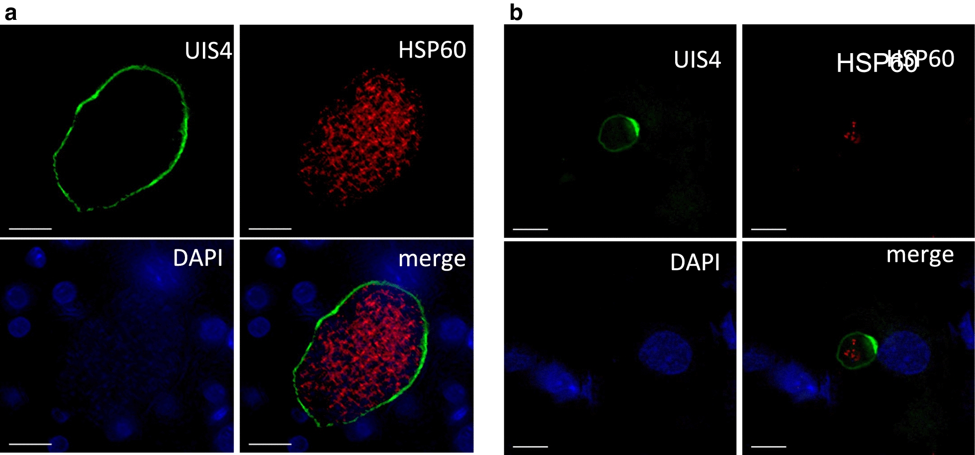
Fig. 4.

α-rUIS4 mAb staining in vivo. Liver-chimeric FRGN huHep mice were infected intravenously with one million P. vivax sporozoites. On day 8 post infection, livers were harvested, fixed and stained with α-rUIS4 mAb and the parasite mitochondrial marker anti-HSP60 as described above. DNA was stained with DAPI. a Representative image of a schizont in which α-rUIS4 mAb continuously stains the PVM. b Representative image of a hypnozoite in which the UIS4-positive prominence is clearly visible. Scale bars = 10 μm
