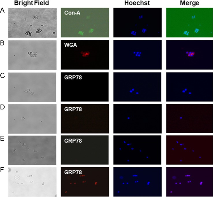
Fig. 4.
Localization of GRP78 in ER–/PR–/HER2– (triple negative; MBA-MB-231) breast cancer cells. Cells were cultured overnight, removed by nonenzymatic cell dissociation solution. The surface N-glycans were stained with (A) FITC-conjugated Con-A (green, 20×), and (B) Texas-red conjugated WGA (red; 20×) in a 50 mM Tris buffer, pH 7.0 containing 0.15 M NaCl and 4 mM CaCl2 but no fixative. The images were collected by a fluorescence microscope (Axioskop 2, Carl Zeiss, Germany) with AxioCam MRc5 camera and Axion Vision Rel 4.6 software. (C–E) Detection of GRP78 (red, 40×); (C) cells were cultured for 24 h in EMEM with 10% serum and then stained with anti-GRP78 antibody; (D) cells were cultured for 24 h in EMEM without serum (i.e., serum-free; 0% FBS) and then stained with anti-GRP78 antibody; (E) cells were treated with tunicamycin (1 μg/mL) for 24 h in EMEM containing 2% serum and the stained with anti-GRP78 antibody; (F) cells were treated with tunicamycin (1 μg/mL) as in (E) and then permeabilized by exposing to ice-cold methanol for 15 s and stained with anti-GRP78 antibody.

