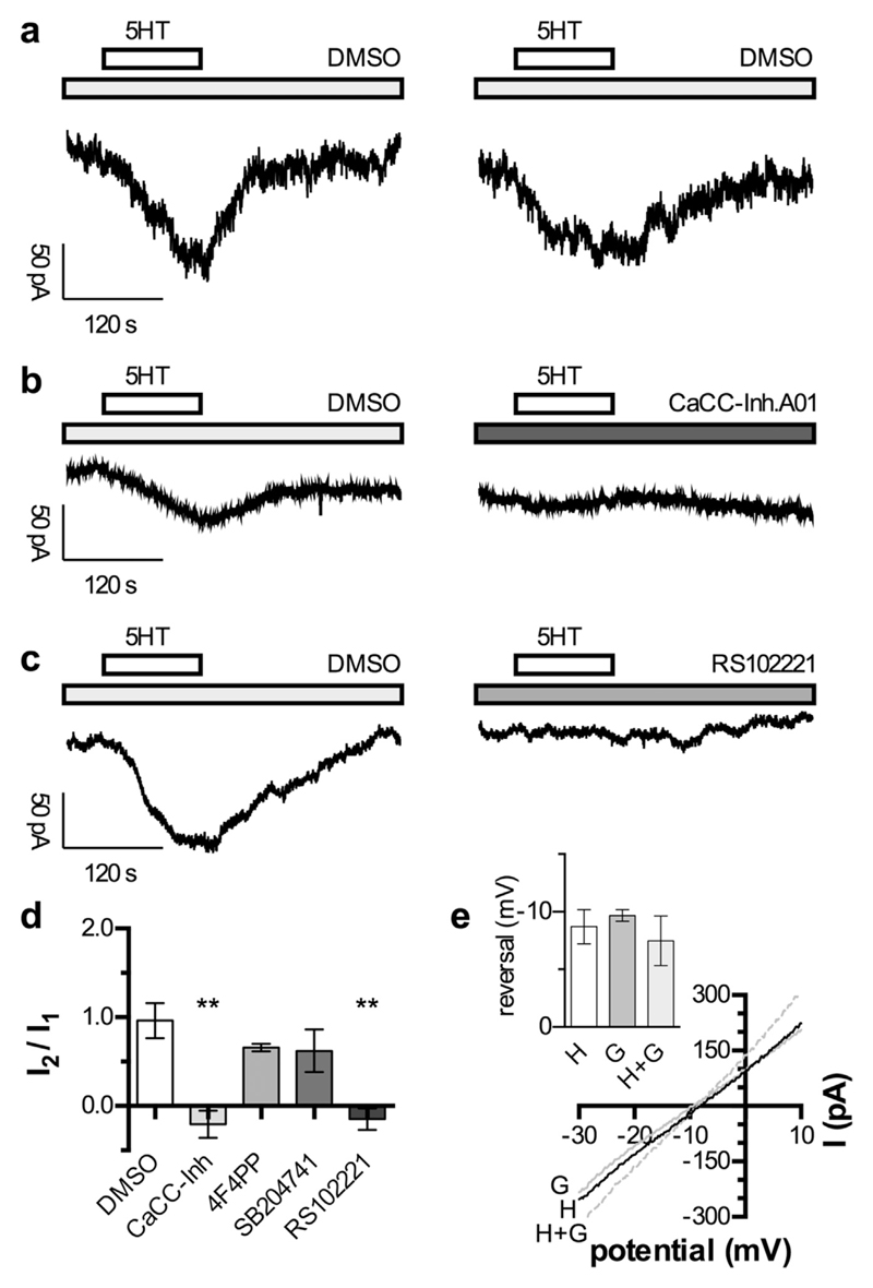
Fig. 4. Activation of currents through CaCCs channels by 5HT.
Cells were recorded in perforated voltage clamp mode at a holding potential of −65 mV (a to d). 5HT was applied twice for 120 s (indicated by the bar) separated by 300 s washout and 60 s wash-in of the respective modulator. a. Original traces of currents induced by two subsequent applications of 10 μM 5HT in presence of 0.03% DMSO. b. Original traces of currents induced by two subsequent applications of 10 μM 5HT in the presence of 0.03% DMSO and 3 μM CaCC-Inh. A01 (as indicated by the grey bar), respectively. c. Original traces of currents induced by two subsequent applications of 10 μM 5HT in the presence of 0.03% DMSO and 30 nM RS102221 (as indicated by the grey bar), respectively. d. Relation of the currents induced by two subsequent applications of 10 μM 5HT (I2/I1) in the presence of 0.03% DMSO (n = 5), 3 μM CaCC-Inh. A01 (n = 5), 100 nM 4F4PP (n = 5), 200 nM SB204741 (n = 5), and 30 nM RS102221 (n = 5), respectively. ** indicate significant differences vs. the I2/I1 ratio obtained in DMSO at p < 0.01, respectively (Kruskal-Wallis test, followed by Dunn's multiple comparison). e. Currents were evoked by the application of 10 μM 5HT (H), 10 μM GABA (G), or 10 μM 5HT plus 10 μM GABA (H + G). During peak current responses, 50 ms ramp depolarizations from −30 mV to +10 mV were applied in order to determine reversal potentials, and the resulting current traces are shown. In the inset, the values of reversal potentials obtained with 10 μM 5HT (H), 10 μM GABA (G), or 10 μM 5HT plus 10 μM GABA (H + G) in 5 different neurons are indicated. There were no significant differences between these three values (p > 0.05; Kruskal-Wallis test, followed by Dunn's multiple comparison).

