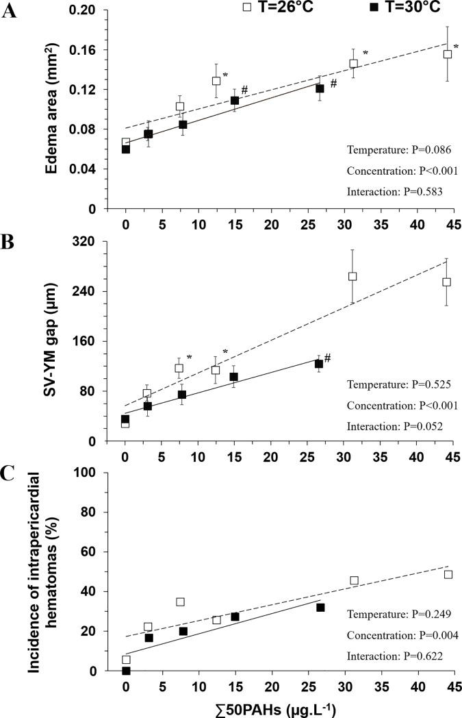
Fig 2. Morphological impairments associated with edema.
(A) Edema area (mm2), (B) sinus venosus-yolk mass gap (μm) and (C) total incidence of intrapericardial hematomas measured at two rearing temperatures 26°C (N = 20–39) and 30°C (N = 20–56). Data for edema area and SV-YM gap are expressed as mean±SEM. Incidence of intrapericadial hematomas is expressed in total percent of hematomas scored in oil—exposed individuals. Simple linear regressions are given for graphical representation at both exposure temperatures. * (26°C) and # (30°C) indicate significant differences of oil exposure concentrations compared to respective control groups (P<0.05).

