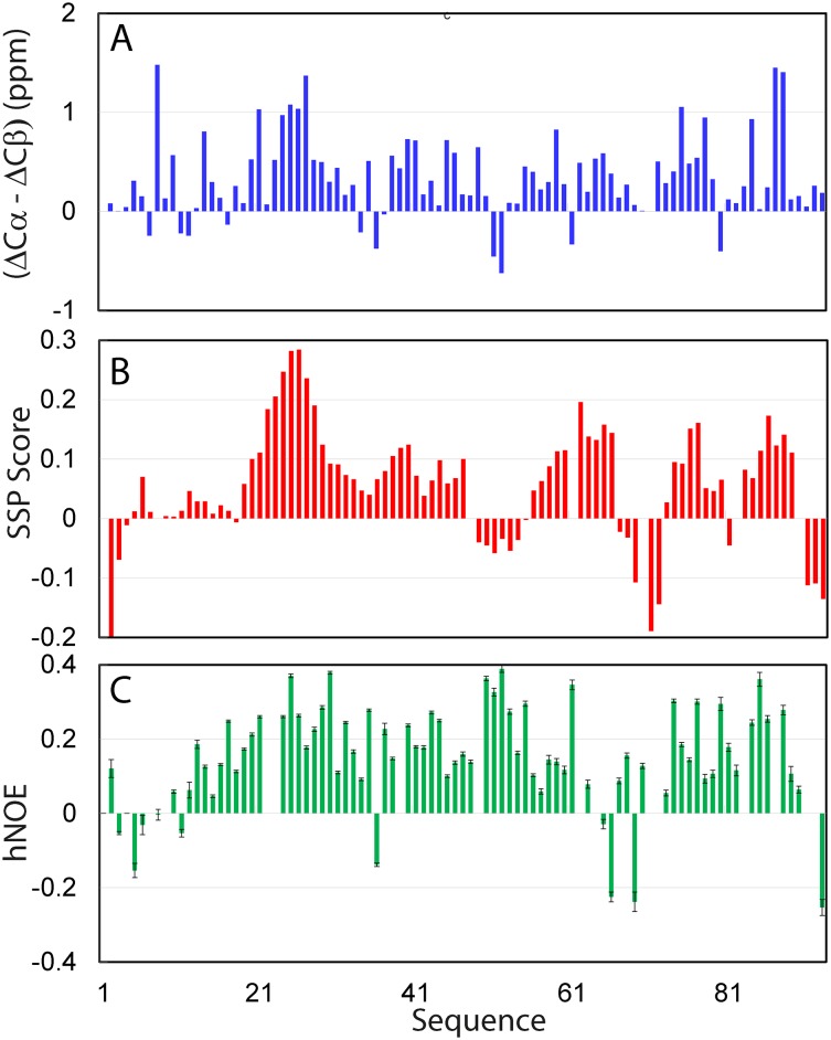
Fig 3. Residue-specific conformation of the TMEM106B cytoplasmic domain.
(A) Residue specific (ΔCα-ΔCβ) chemical shifts of the TMEM106B cytoplasmic domain. (B) Secondary structure propensity (SSP) score obtained by analyzing chemical shifts of the TMEM106B cytoplasmic domain with the SSP program. A score of +1 is for the well-formed helix while a score of -1 for the well-formed extended strand. (C) {1H}-15N heteronuclear steady-state NOE (hNOE) of the TMEM106B cytoplasmic domain.

