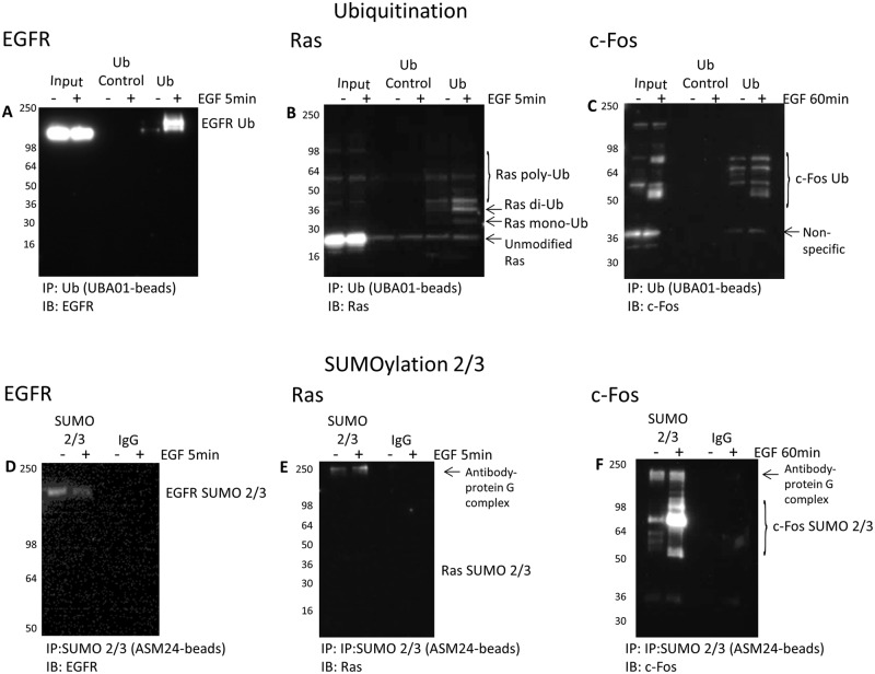
Figure 6. Characterize Ub and SUMO 2/3 for the EGFR–Ras-c–Fos axis.
Serum-restricted A431 cells were either unstimulated or stimulated with EGF for 5 or 60 min prior to lysis with BlastR lysis buffer. (A–C) Samples were immunoprecipitated with ubiquitin control beads (CUB02) or ubiquitin-binding beads (UBA01). Samples were separated by SDS/PAGE and analyzed by Western blot for (A) EGFR, (B) Ras, and (C) c-Fos to identify the ubiquitinated species for these proteins in the EGFR signaling pathway. Shown are representative Western blots from n≥3 independent experiments. (D–F) Samples were immunoprecipitated with IgG control beads or SUMO 2/3 binding beads (ASM24). Samples were separated by SDS/PAGE and analyzed by Western blot for (D) EGFR, (E) Ras, and (F) c-Fos to identify the SUMOylated 2/3 species for these proteins in the EGFR signaling pathway. Shown are representative Western blots from n≥3 independent experiments.

