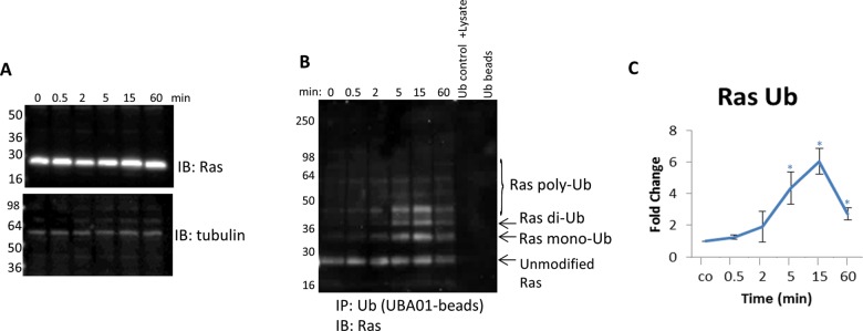
Figure 7. Detect endogenous, temporal changes of Ras Ub in response to EGF stimulation.
(A) Serum-restricted A431 cells were stimulated with EGF for the given time period. Whole cell lysate (WCL) was analyzed for Ras levels. Tubulin was used as a loading control. (B) Unstimulated and EGF-treated A431 lysates were incubated with ubiquitin-binding beads (UBA01) to immunoprecipitate ubiquitinated proteins or ubiquitin control beads (CUB02). Samples were separated by SDS/PAGE and analyzed by Western immunoblotting using a pan Ras antibody to identify ubiquitinated pan Ras. Shown are representative Western blots from n≥3 independent experiments. (C) Quantitation of densitometric analysis of endogenous ubiquitinated Ras in response to EGF stimulation. Error bars represent S.E.M. t test statistical analysis was performed. *P<0.05.

