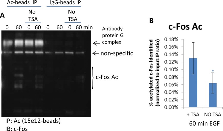
Figure 8. Identify and validate c-Fos Ac.
(A) Untreated or 60 min EGF-treated A431 cells were harvested with BlastR lysis buffer with or without TSA. Lysates were incubated with acetyl lysine binding beads or IgG control binding beads. Samples were separated by SDS/PAGE and analyzed by Western blot for acetylated c-Fos with a c-Fos antibody; shown are representative Western blots from n≥3 independent experiments. (B) Quantitation of densitometric analysis of c-Fos Ac from lysates with or without TSA. Samples were normalized to total c-Fos, as well as, input: IP ratio (0.008). Error bars represent S.E.M. t test statistical analysis was performed. *P<0.05.

