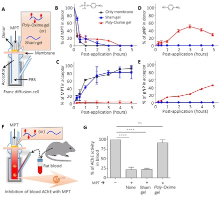
Fig. 2. Poly-Oxime gel deactivates MPT to limit MPT-mediated AChE inhibition ex vivo.

(A) Schematic of Franz diffusion cell: A thin layer of either poly-Oxime gel or sham gel was applied on a dialysis membrane, which was placed between donor and acceptor chambers. (B to E) Concentration of MPT and pNP (hydrolytic degradation product of MPT) in donor and acceptor chambers was measured using UFLC. The presence of sham gel did not prevent the diffusion of MPT into the acceptor chamber and could not hydrolyze MPT to generate pNP, whereas poly-Oxime actively hydrolyzed to limit the penetration of toxic MPT into the acceptor chamber. (F and G) An ex vivo assay to demonstrate the ability of poly-Oxime to limit MPT-induced assay AChE inhibition using rat blood. AChE containing rat blood was placed in the acceptor chamber, and MPT was added in the donor chamber in the presence of either poly-Oxime or sham gel. Active AChE was measured in the blood before and 3 hours after addition of MPT. In the absence of poly-Oxime gel, MPT diffused into an acceptor chamber and significantly inhibited AChE activity. However, poly-Oxime gel could hydrolyze MPT before diffusion, therefore limiting the MPT-induced inhibition of AChE. Data are means ± SD (n = 3, performed at least twice); P values were determined by one-way analysis of variance (ANOVA). ****P < 0.0001. ns, not significant.
