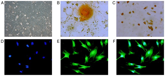
Figure 1.
Identification of BMSC morphology and differentiation functions. (A) BMSCs were observed under a microscope at magnification ×100. (B) BMSCs cultured in osteogenic medium for 3 weeks. Dark brown calcium nodules stained by alizarin red were observed at magnification, ×400. (C) BMSCs cultured in adipogenic medium for 3 weeks. Brown lipid droplets stained by oil red O were observed at magnification, ×400. BMSCs-GFP cells exhibited (D) blue nuclei stained by DAPI and (E) GFP labeled actin cytoskeleton under a fluorescence microscope at magnification, ×400. (F) Merged of (D) and (E) indicates 100% overlap between GFP (green) and DAPI (blue). BMSC, bone mesenchymal stem cells; GFP, green fluorescence protein.

