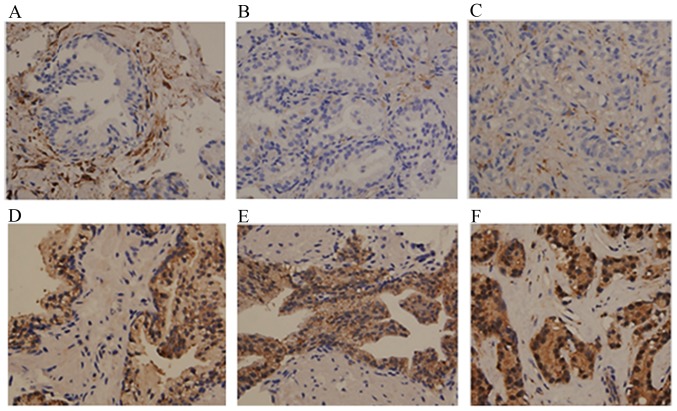
Figure 2.
Immunohistochemical staining of PKC-α and PKC-ι in BPH, HGPIN and PC. Tissue was stained with PKC-α antibody as a control for PKC-ι staining. Results show PKC-ι staining in (A) BPH, (B) HGPIN, and (C) PC tissue. PKC-α staining is shown in (D) BPH glands, (E) glands with HGPIN, and (F) PC glands. Tissues examined comprised BPH (n=9), HGPIN (n=8) and PC tissues (n=10). Magnification for all micrographs was x400. Three experiments were performed in triplicate. BPH, benign prostate hyperplasia; HGPIN, high grade prostate intraepithelial neoplasma; PC, prostate adenocarcinoma; PKC, protein kinase C.

