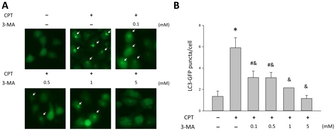
Figure 5.
3-MA treatment suppresses CPT-induced accumulation of autophagic markers in H1299 cells. (A) White arrows indicate GFP-LC3 puncta in H1299 cells following treatment with various concentrations of 3-MA, in the presence of 0.5 µM CPT. Magnification, ×200. (B) GFP-LC3 puncta/cell was observed under a microscope and counted. Data are presented as the means ± standard deviation. #P<0.05 vs. the control group; *P<0.001 vs. the control group; &P<0.001 vs. the CPT-treated group. 3-MA, 3-methyladenine; CPT, camptothecin; GFP, green fluorescent protein; LC3, microtubule-associated proteins 1A/1B light chain 3.

