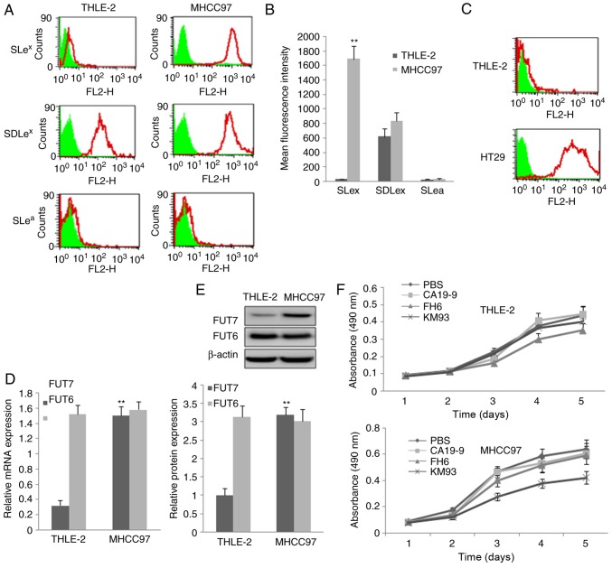
Figure 1.
Expression of three sialyl-Lewis oligosaccharides and relative FUTs and the inhibition effect of monoclonal antibody on cell proliferation. (A) MHCC97 human hepatocellular carcinoma cells and THLE-2 normal liver cells were assayed by flow cytometry using three monoclonal antibodies targeting sialyl-Lewis antigens. PBS, cells treated with PBS and without antibodies as a control; KM93, cells treated with mAb against SLex; FH6, cells treated with mAb against SDLex; CA19-9, cells treated with mAb against SLea. In the flow cytometry histograms, the areas in green show the number of unstained cells and the areas outlined in red represent cells binding to mAb. (B) Quantitative analysis of the expression of sialyl-Lewis oligosaccharides by calculation of mean fluorescence intensity. THLE-2 cells were used as a control. (C) THLE-2 cells and HT29 cells were assayed by flow cytometry using monoclonal antibody against SLea antigen to confirm the high affinity of the SLea antibody. (D) mRNA expression of FUT7 and FUT6 were detected by reverse transcription-quantitative polymerase chain reaction analysis. The relative gene mRNA level was normalized to the respective glyceraldehyde-3-phosphate dehydrogenase level. THLE-2 cells were used asa control. (E) Protein expression levels of FUT7 and FUT6 protein were evaluated by western blot analysis (above) and quantitative analysis of relative protein levels was performed (below). THLE-2 cells were used as a control. **P<0.01 (n=3), vs. THLE-2 Control. (F) Effect of monoclonal antibodies on the proliferation of THLE-2 cells and MHCC97 cells. The 3-(4,5-dimethylthiazol-2-yl)-2,5-diphenyl-2H tetrazolium bromide assay showed that MHCC97 cell proliferation was significantly suppressed by KM93, a mAb against SLex. FUT, fucosyltransferase; SLex, sialyl-Lewis X, SLea, sialyl-Lewis a; SDLe X, dimeric SLe; mAb, monoclonal antibody.

