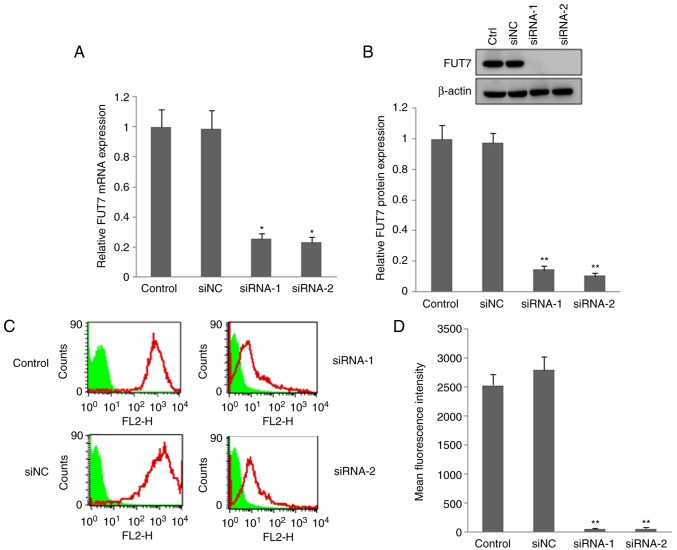
Figure 2.
Changes in the expression of FUT7 and SLex of MHCC97 cells following transfection with FUT7 siRNAs. siRNA targeting FUT7 was transfected into MHCC97 cells and the interference rate was confirmed by RT-qPCR and western blot analyses. Flow-cytometric analysis was used to detect the expression of SLex. (A) mRNA level of FUT7 detected by RT-qPCR analysis following FUT7-siRNA transfection. (B) Protein level of FUT7 detected by western blot analysis following FUT7-siRNA transfection (above) and quantitative analysis of the protein level of FUT7 following FUT7-siRNA transfection below). (C) Flow cytometric analysis of cell surface expression of SLex following FUT7-siRNA transfection. (D) Quantitative analysis of the expression of SLex by calculating the mean fluorescence intensity. *P<0.05 (n=3), vs. untransfected control cells; **P<0.01 (n=3), vs. untransfected control cells. FUT, fucosyltransferase; SLex, sialyl-Lewis X; RT-qPCR, reverse transcription-quantitative polymerase chain reaction; siRNA, small interfering RNA; siNC, scramble-siRNA transfected cells; Control/Ctrl, untransfected cells.

