Figure 5.
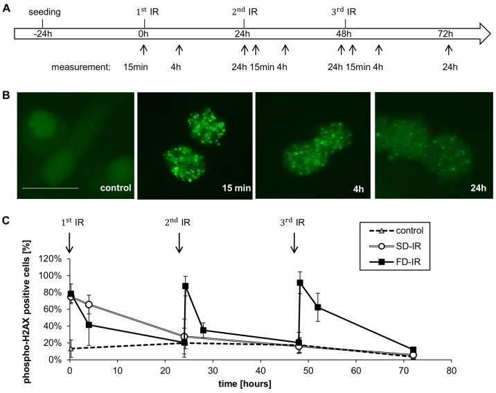
Different DNA damage response after high single-dose irradiation (SD-IR) and fractionated dose irradiation (FD-IR). (A) Experimental chronology: G35 DCs were seeded 24 h before the first radiation and re-irradiated every 24 h in the FD-IR group, or mock treated in the SD-IR group. Irradiation was delivered either once (SD-IR 1x6 Gy) or in fractions (FD-IR 3x2 Gy). (B) Cells were fixed in formaldehyde and immunohistochemically stained with phospho- H2AX, as surrogate marker for a DNA damage response. and examined with a fluorescence microscope. Representative images of phospho-H2AX stained cells 15 min, 4 and 24 h after 2 Gy single dose irradiation are shown. Scale bar, 25 µm. (C) The percentage of phospho-H2AX-positive cells was determined after counting cells 15 min, 4 and 24 h after (mock) irradiation. Cells with >10 foci per nucleus were considered positive. Shown is the means ± SD of three independent experiments.

