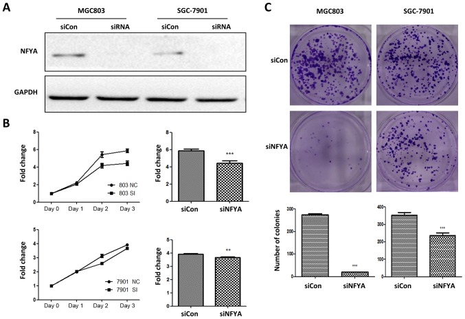
Figure 7.
Investigation of the role of NFYA in diffuse and intestinal-derived GC cells. (A) Western blot analysis of NFYA knockdown by siRNA in MGC803 and SGC-7901 cells. (B) CCK-8 assay of MGC803 (upper panel) and SGC-7901 (lower panel) cell growth in the presence of siNFYA; the growth curve is presented on the left, and the fold change at day 3 is presented on the right. (C) Colony formation assays of MGC803 (left panel) and SGC-7901 (right panel) in the presence of siNFYA; representative colony formation results are presented in the upper panel, and the statistical results are presented in the lower panel. All assays were performed in triplicate. **P<0.01; ***P<0.001. NFYA, nuclear transcription factor Y subunit α; siCon, control siRNA; siNFYA, NFYA siRNA; siRNA, small interfering RNA.

