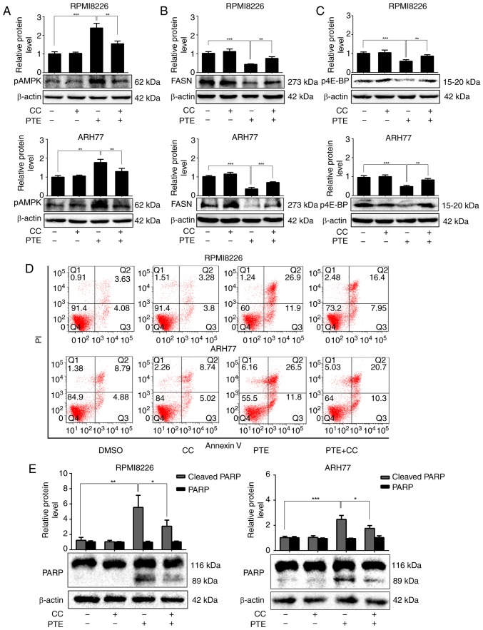
Figure 3.
PTE induces MM cells apoptosis in a AMPK-dependent manner. RPMI-8226 and ARH-77 cells were treated with PTE in the absence or presence of CC. Western blotting analyzed the levels of (A) p-AMPK (Thr 172), (B) FASN and (C) p-4E-BP1. β-actin served as a loading control; (D) Flow cytometric analysis of apoptotic effects using an Annexin V-FITC/PI kit. (E) Western blot analysis of the levels of PARP. β-actin served as a loading control. *P<0.05, **P<0.01, ***P<0.001 vs. PTE-only group. PTE, pterostilbene; MM, multiple myeloma; AMPK, AMP-activated protein kinase; CC, compound C; p, phosphorylated; FASN, fatty acid synthase; 4E-BP1, 4E-binding protein 1; FITC, fluorescein isothiocyanate; PARP, poly(ADP-ribose) polymerase.

