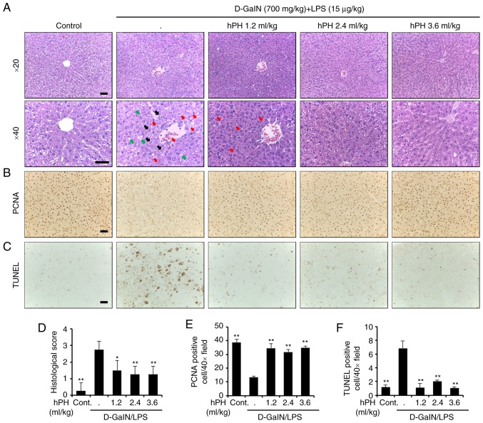
Figure 2.
Effects of hPH treatment on D-GalN/LPS-induced acute liver failure in tissue degeneration and apoptosis. (A) Histopathological staining. Upper panel (magnification, ×20), lower panel (magnification, ×40). Scale bar=100 µm. The arrows indicate apoptotic hepatocytes (black), a severely affected region of a lobule containing ballooned hepatocytes (red), and hepatocytes expanded by fat vacuoles (green). (B) Images showing PCNA staining. Scale bar=100 µm. (C) Apoptotic response to D-GalN/LPS in the liver tissue of rats was investigated using TUNEL staining. Scale bar=100 µm. (D) Stained sections were graded for histopathology using a four-point scale (0-3), with 0, 1, 2, and 3 representing no damage, mild damage, moderate damage, and severe damage, respectively. (E) PCNA-positive cells were significantly less numerous in the D-GalN/LPS-treated group than in the control group; the addition of hPH led to significant overexpression of PCNA in the liver compared with D-GalN/LPS treatment alone. (F) TUNEL-positive cells was higher in number in the group treated with D-GalN/LPS alone compared with the control group. However, apoptotic induction by D-GalN/LPS was further reduced in the groups treated with 1.2, 2.4, and 3.6 ml/kg of hPH. All data are presented as the mean ± standard error of the mean. *P<0.05 and **P<0.01, vs. D-GalN/LPS group. hPH, human placental hydrolysate; D-GalN, D-galactosamine; LPS, lipopolysaccharide; PCNA, proliferating cell nuclear antigen; Cont, control.

