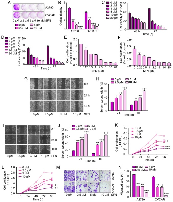
Figure 1.
Sulforaphane effectively suppresses human ovarian cancer cell proliferation. (A) Colony formation assay. Subconfluent A2780 and OVCAR cells were seeded in plates and treated with sulforaphane at the concentrations shown. After 48 h, the cancer cells were fixed and stained with crystal violet for the colony formation assay. (B) Colony-forming ability of ovarian cancer cell lines following sulforaphane treatment was quantified for optical absorbance. Cell viability assay. (C) A2780 and (D) OVCAR cells were seeded in plates and treated with sulforaphane at the concentrations shown. After 48 and 72 h, the viable cells were collected, stained and counted under a bright field microscope. WST-1 cell proliferation assay. (E) A2780 and (F) OVCAR cancer cells were seeded in 96-well plates and treated with sulforaphane at the indicated concentrations. At 24 h post-treatment, WST-1 reagent was added to plates and incubated for 1 h, and absorbance measurement was performed. All assay conditions were performed in triplicate. Cell wounding analysis of (G) A2780 cells with (H) quantification, and (I) OVCAR cells with (J) quantification. Cells were wounded with the micro-pipette tips and treated with sulforaphane at the indicated concentrations. The wound gap was measured at 0, 24 and 48 h post-sulforaphane treatments (magnification, ×40). The proliferation assay indicated that sulforaphane inhibited the proliferation of (K) A2780 and (L) OVCAR ovarian cancer cells. (M) Transwell assay showed the metastasis capacities were inhibited by sulforaphane in A2780 and OVCAR ovarian cancer cells (magnification, ×40). (N) Quantification of the migrated ovarian cancer cells. The values are presented as the mean ± standard deviation (n=8-10) of the samples. +P<0.05, ++P<0.01 and +++P<0.001, vs. 0 μM group. SFN, sulforaphane.

