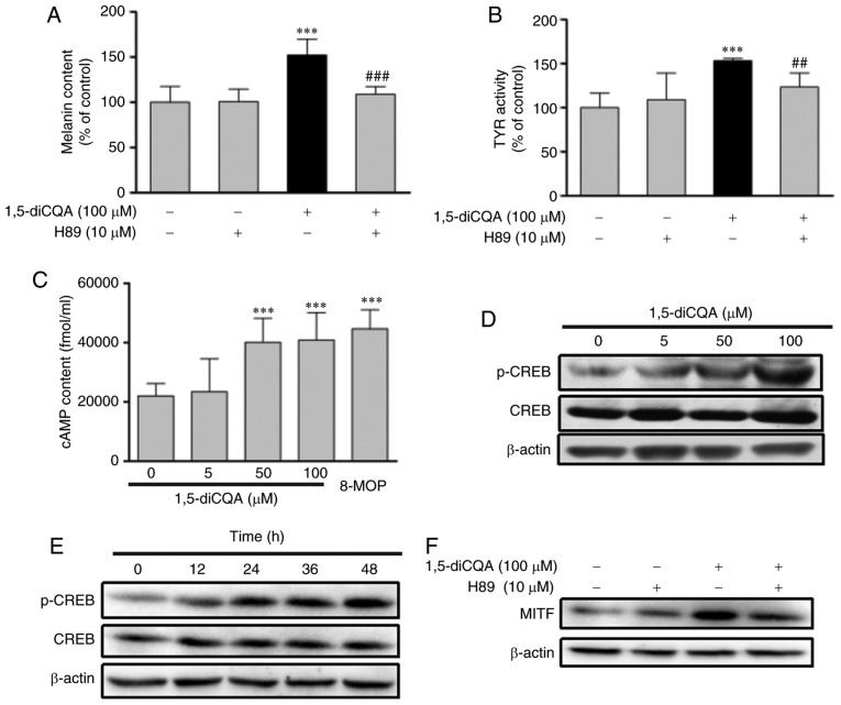
Figure 9.
Effects of 1,5-diCQA on the PKA signal pathway. B16 cells were pre-incubated with H89 (10 µM) for 2 h prior to the addition of 1, 5-diCQA (100 µM), and then (A) incubated for 48 h for the measurement of melanin content or (B) incubated for 24 h for the measurement of TYR activity. (C) B16 cells were treated with 0.1% dimethyl sulfoxide and 8-MOP as positive controls or 1,5-diCQA at 5, 50 and 100 µM for 12 h, and then cAMP content was measured by a cAMP-ELISA kit. (D) B16 cells were treated with 5, 50 and 100 µM 1,5-diCQA for 48 h, and levels of total and phosphorylated CREB were measured by western blot analysis. (E) B16 cells were treated with 1,5-diCQA (100 µM) for 0, 12, 24, 36 and 48 h, and levels of total and phosphorylated CREB were measured by western blot analysis. (F) B16 cells were pre-incubated with H89 (10 µM) for 2 h prior to the addition of 1,5-diCQA (100 µM), and then incubated for 48 h. MITF expression levels were the measured by western blot analysis. **P<0.01 and ***P<0.001 vs. untreated control group. ##P<0.01 and ###P<0.001 vs. single treatment group. PKA, protein kinase A; TYR, tyrosinase; MITF, microphthalmia-associated transcription factor; 1,5-diCQA, 1,5-dicaffeoylquinic acid; p-phosphorylated; CREB, cAMP-response element binding protein.

