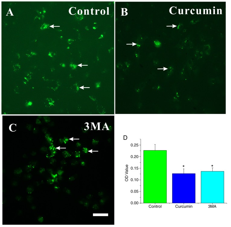
Figure 5.
MDC-labeled AVOs occur in curcumin- and 3MA treated NSCs. Characterization of the MDC-labeled vacuoles. NSCs were seeded on slides. Cells were incubated in proliferation medium and (A) left untreated or treated with 10 µM (B) curcumin or (C) 3MA for 72 h, and then stained with 0.05 mM MDC. (D) Cells were mounted and analyzed by fluorescence microscopy. The fluorescence intensity values (OD value) of all groups were digitally quantified .using ImageJ. The results are presented as the mean ± standard deviation (n=3). Scale bar, 25 µm. *P<0.05 compared with control. MDC, monodansylcadaverine; AVO, autophagic vacuoles; 3MA, 3-methyladenine; NSC, neural stem cells.

