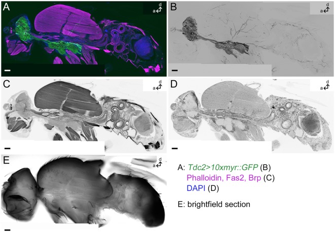
Figure 1.
Tdc2-Gal4-positive arborizations in a whole fly section. Projection of one medial sagittal agarose section of 80 µm thickness labeled by anti-GFP to visualize membranes of Tdc2-Gal4-positive neurons (green in (A), black in (B)); Phalloidin, anti-Fasciclin2 (Fas2) and anti-Bruchpilot (Brp) to visualize muscles, cells and synapses, respectively (magenta in (A), black in (C)) and DAPI to mark cell bodies (blue in (A), black in (D)). (E) A single optical section showing the bright-field picture. Scale bars = 50 µm.

