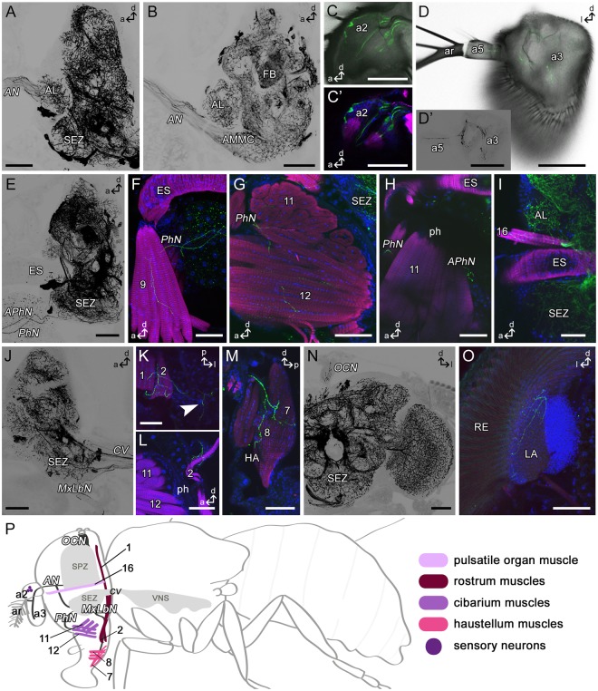
Figure 2.
Tdc2-Gal4-positive arborizations in the fly’s head. Projections of sagittal (A–C,E–J,L-M) or frontal (D,N-O) or horizontal (K) optical sections visualizing the arborization pattern of Tdc2-Gal4-positive neurons (Tdc2N; black or green) in the head. (A–D) Tdc2Ns run through the antennal nerve (AN) and project in antennal segments a2, a3 and a5. Mechanosensory neurons of the Johnston’s organ are visible (C). (E–H) Efferent Tdc2Ns of the pharyngeal (PhN) and accessory pharyngeal nerve (APhN). (F,G) Cells of the PhN innervate muscles 9, 11 and 12. (H) Bouton-like structures of APhN neurons beside the pharynx (ph). (I) Innervation of muscle 16. (J–M) Tdc2Ns of the maxillary-labial nerve (MxLbN) project along muscle 1 and 2 (K,L) in the haustellum (HA) and seem to innervate muscles 7 and 8 (M). Arborizations from the MxLbN reach the lateral brain (arrowhead in K). (N) Tdc2Ns arborize in the ocellar nerve (OCN). (O) Ramifications in the lateral lamina (LA) close to the retina (RE). (P) Schematic drawing of a fly visualizing peripheral nerves (dark grey), muscles of the head and proboscis (rose to pink) and sensory neurons (purple) shown in A-N. a, anterior; AL, antennal lobe; AMMC, antennal mechanosensory and motor center; ar, arista; CV, cervical connective; d, dorsal; ES, esophagus; FB, fan-shaped body; l, lateral; p, posterior; SEZ, subesophageal zone. Scale bars = 50 µm.

