Figure 3.
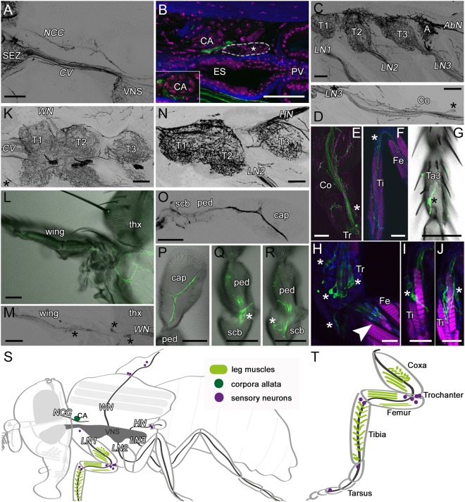
Tdc2-Gal4-positive arborizations in the thorax. Projections of optical sections visualizing the arborization pattern of Tdc2-Gal4-positive (Tdc2Ns; A–N,Q,R) and Tdc2-positive (O,P) neurons (black or green) in the thorax. (A) Tdc2Ns run through the cervical connective (CV) and corpora cardiaca nerve (NCC). (B) Tdc2Ns arborize close to the corpora allata (CA) and the anterior stomatogastric ganglion (white-rimmed). (C–F) Tdc2Ns project along the legs and innervate leg muscles. (G) An afferent sensory neuron in the third segment of the tarsus (Ta). (H–J) Cell bodies of sensory neurons (asterisks) of the trochanter (Tr; H) and tibia (Ti; I,J). (H) Neurons of the chordotonal organ in the femur (Fe; arrowhead). (K–M) Tdc2Ns project along the wing nerve (WN). (K) Innervation of the thoracic chordotonal organ (asterisk). (L,M) Tdc2Ns run along the L1 wing vein. Cell bodies of sensory neurons are visible (asterisks). (N–R) Tdc2Ns in the haltere nerve (HN). (O,P) anti-Tdc2-positive cells project to the distal part of the capitellum (cap). (Q,R) Sensory neurons of the pedicellus (ped) and scabellum (scb) are labeled by Tdc2-Gal4 and Tdc2 antibody (O). (S,T) Schematic drawing of a fly and one leg visualizing the VNS and peripheral nerves (dark grey), CA (dark green), leg muscles (light green) and sensory neurons (purple) shown in A–R. (A–J,N–R) sagittal sections; (K) horizontal sections; (L,M) frontal sections. A, abdominal segment; Co, coxa; ES, esophagus; LN, leg nerve; PV, proventriculus; SEZ, subesophageal zone; T1–3, thorax segment1–3; thx, thorax. Scale bars: A–G,K–R = 50 µm; H–J = 25 µm.

