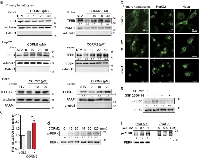
Fig. 1. Carbon monoxide (CO) increases TFEB nuclear translocation.
a Primary hepatocytes, HepG2, or HeLa cells were incubated with CORM2 at the indicated doses or starved (STV) for 3 h. Cells were fractionated into cytosol and nuclear. Endogenous TFEB proteins were analyzed by immunoblotting. HeLa cells were transfected with TFEB-GFP plasmid for 24 h and then treated with CORM2 at the indicated dose for 3 h. Nuclear translocation of CORM2-induced TFEB-GFP was detected by anti-GFP antibody. b Primary hepatocytes, HepG2, and HeLa cells were transfected with TFEB-GFP plasmid for 24 h. After transfection, cells were treated with CORM2 (20 μM) or Torin 1 (1 μM) for 3 h and TFEB localization was determined by confocal fluorescence microscopy. Scale bar, 20 μm. c Primary hepatocytes were co-transfected with 4X CLEAR-luciferase reporter construct and pRL-SV40 Renilla luciferase construct for 24 h. After treatment with CORM2 for 9 h, cells were lysed and assayed for luciferase activity. Values are shown as mean ± SEM (n = 3). ***P < 0.001. d Primary hepatocytes were treated with CORM2 (20 μM) for the indicated times. The levels of phosphorylated PERK (p-PERK) and total PERK were measured by immunoblotting. e Primary hepatocytes were pretreated with GSK2606414 (PERK inhibitor, 0.5 μM) for 30 min and then treated with CORM2 (20 μM) for 1 h. The levels of phosphorylated PERK (p-PERK) and total PERK were measured by immunoblotting. f Perk+/+ and Perk−/− MEF cells were treated with CORM2 (20 μM) for 30 min and 1 h. The levels of phosphorylated PERK (p-PERK) and total PERK were measured by immunoblotting. Band intensities were determined by densitometry (Image J). Basal levels in the untreated sample were set at 1.0 and results are expressed as fold induction over control levels

