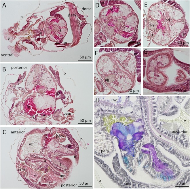
Figure 3.
Histological observation of Crassostrea gigas pediveliger larvae stained with hematoxylin and eosin (A–G) and multichrome technic (Alcian Blue, Periodic Acid–Schiff’s, Haematoxylin, Picric Acid) (H). A: Transverse, B: Frontal, C and H: Sagittal sections of the whole larvae. (D–G) show serial transverse sections of a foot. aam: anterior adductor muscle, bd: byssus duct, f: foot, fg: foot gland, fm: foot muscle, gr: gill rudiment, m: mantle, mo: mouth, o- oesophagus, p: periostracum, pam: posterior adductor muscle, pg: pedal ganglia, v: velum, vc: visceral cavity, arrow: secreting cells from the foot gland.

