Fig. 6.
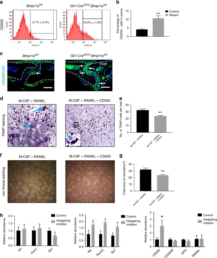
Upregulated CD200 may contribute to decreased osteoclastogenic activity in Gli1-CreERT2;Bmpr1afl/fl sutures. a FACS analysis of CD200 in suture mesenchymal cells collected from Bmpr1αfl/fl (control) and Gli1-CreERT2;Bmpr1αfl/fl (mutant) mice 2 weeks post induction. b Quantitation of the percentages of CD200+ cells in suture mesenchyme from three independent samples. c Immunostaining of CD200 (green, indicated by arrows) in the suture mesenchyme of Bmpr1αfl/fl (control) and Gli1-CreERT2;Bmpr1αfl/fl (mutant) mice 2 weeks post induction (2wpt). Broken lines indicate the outline of the suture. d TRAP staining of osteoclasts induced from BMMs of 4-week-old C57BL/6J mice, after culture with M-CSF for 3 days and then with RANKL or RANKL plus exogenous CD200 for another 5 days. The inset shows a magnified view of the boxed region. Blue arrows indicate mature osteoclasts. e Quantitation of multinucleated TRAP+ osteoclasts per well in four independent experiments. f Resorption activity assay (von Kossa staining) of osteoclasts induced from BMMs of 4-week-old C57BL/6J mice, after culture with M-CSF for 3 days and then with RANKL or RANKL plus exogenous CD200 for another 5 days. g Quantitation of the percentage of resorption in four independent experiments. h Real-time PCR of hedgehog signalling members (Ihh, Ptch1, and Gli1), osteogenic markers (Alp, Runx2, and Sp7), and osteoclastic-related markers (CD200, CD200R, OPG, and RANKL) in the suture mesenchyme of C57BL/6J mice 2 weeks post treatment with or without GDC0449 (hedgehog inhibitor) from five independent samples. T tests were performed. *P < 0.05; **P < 0.01. Scale bars, 100 µm

