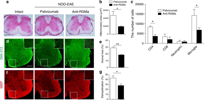
Fig. 4. Treatment with humanized anti-RGMa antibody suppresses inflammation, demyelination, and neurodegeneration in NOD-EAE mice.
a Lumbar spinal cord sections from control mice and NOD-EAE mice treated with palivizumab (EAE score: 4) or anti-RGMa antibody (EAE score: 2) were evaluated by HE staining at 70 days of immunization. Scale bar: 200 μm. Images are representative of spinal cords extracted from at least three mice per treatment group. b Quantitation of inflammation as assessed in (a) (palivizumab: n = 4, anti-RGMa: n = 4, assessed by Student’s t test). c The number of CD4+ T cells, CD8+ T cells, Ly-6G+ neutrophils, and CD45mid CD11b+ microglia in the spinal cord of NOD-EAE mice treated with palivizumab or anti-RGMa antibody analyzed by flow cytometry at 70 days of immunization (palivizumab: n = 3, anti-RGMa: n = 3, assessed by Student’s t test). d Immunohistochemistry for axonal neurofilaments (SMI-312) from lumbar spinal cord sections of control mice and NOD-EAE mice treated with palivizumab (EAE score: 4) or anti-RGMa antibody (EAE score: 2) at 70 days of immunization. Inset images are a high magnification of the area within the white squares. Scale bar: main image 250 μm; inset 100 μm. e Quantitation of axonal loss as assessed in (d) (palivizumab: n = 4, anti-RGMa: n = 4, assessed by Student’s t test). f Immunohistochemistry for MBP from lumbar spinal cord sections of control and NOD-EAE mice treated with palivizumab (EAE score: 4) or anti-RGMa antibody (EAE score: 2) at 70 days of immunization. g Quantification of demyelination as assessed in (f) (palivizumab: n = 5, anti-RGMa: n = 6, assessed by Student’s t test). Error bars represent mean ± SEM. *p < 0.05, **p < 0.01

