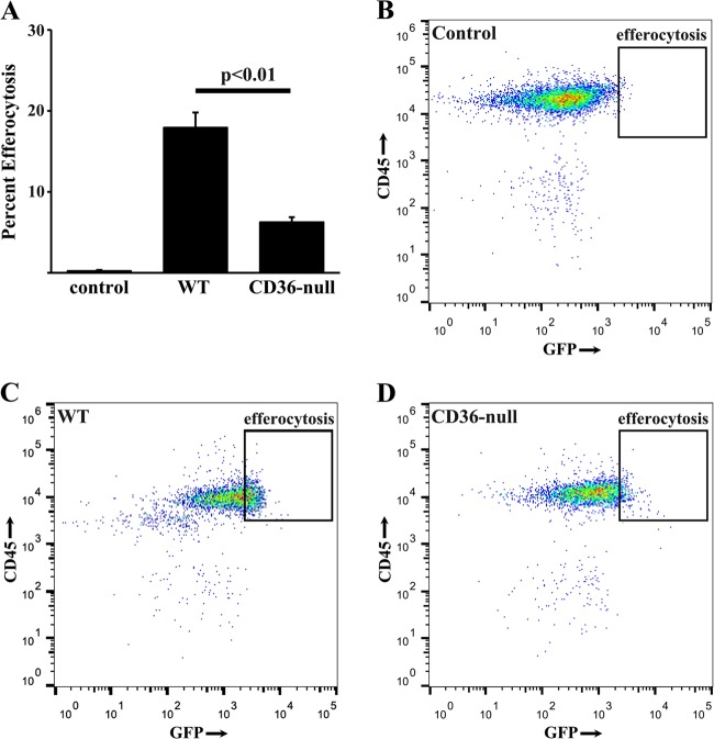
Fig. 6. CD36-null mice exhibit less apoptotic type II AEC efferocytosis.
a BAL cells from WT mice (control) or CD36-null mice 2 h after delivery of PBS or GFP-labeled apoptotic MLE-12 cells were labeded with PE-conjugated anti-mouse CD45 antibody and the percent efferocytosis is quantified by flow cytometry. b-d Representative flow cytometry plots of PBS treated WT control mice (b), UV GFP MLE-12 treated WT mice (c) and UV GFP MLE-12 treated CD36-null mice (d). N = 6 per group

