Figure 1.
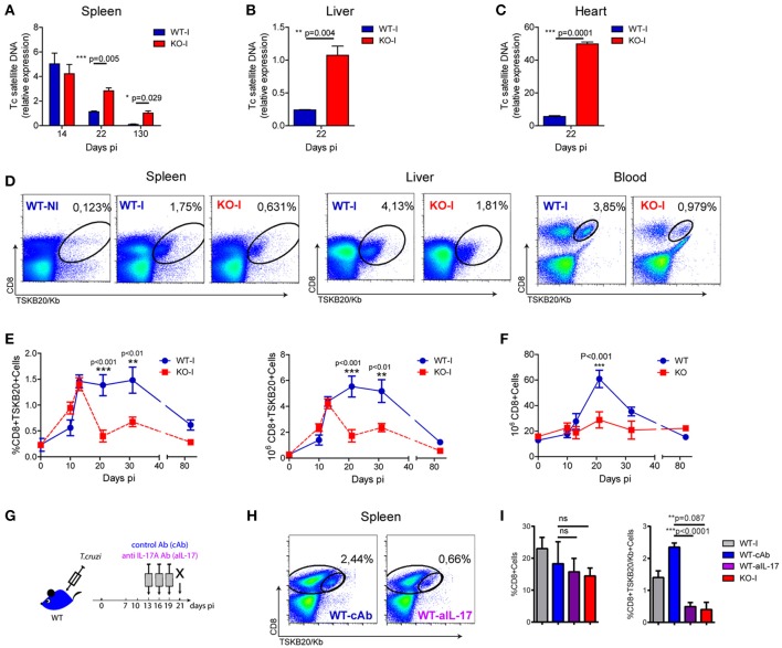
Absence of IL-17RA signaling results in increased tissue parasitism and a reduced magnitude of parasite-specific CD8+ T cell responses. (A-C) Relative amount of T. cruzi satellite DNA in spleen (A), liver (B) and heart (C) of infected WT and IL-17RA KO (KO) mice determined at the indicated dpi. Murine GAPDH was used for normalization. Data are presented as mean ± SD, N = 5 mice. P values calculated with two-tailed T test. (D) Representative plots of CD8 and TSKB20/Kb staining in spleen, liver and blood of WT and KO mice at 22 dpi (WT-I and KO-I, respectively). A representative plot of the staining of splenocytes from non-infected WT mice (WT-N) is shown for comparison. (E) Percentage and cell numbers of TSKB20/Kb+ CD8+ T cells and (F) cell numbers of total CD8+ T cells determined in spleen of WT and IL-17RA KO mice at different dpi. Data shown as mean ± SD, N = 5−8 mice. P values calculated using two-way ANOVA followed by Bonferroni‘s post-test. (G) Experimental layout of IL-17 neutralization in infected WT mice injected with control isotype or anti-IL-17 Abs (WT-cAb and WT-aIL-17, respectively). (H) Representative plots of CD8 and TSKB20/Kb staining in spleen and liver infected control and treated WT mice. (I) Percentage of total and TSKB20/Kb+ CD8+ T cells infected control and treated WT mice. Results from infection-matched WT (WT-I) and KO mice (KO-I) are shown for comparison. Data are representative of five (A–F), and two (G–I) independent experiments.

