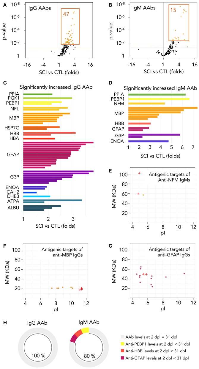
Figure 4.
Autoantibodies against modified central nervous system and peripheral proteins are rapidly increased after injury. (A,B) Among the 173 different IgG and IgM autoantibodies detected, 47 IgGs and 15 IgMs are significantly increased at 1 month after injury (t-test p-value < 0.05 and FDR < 0.05 after multiple comparison correction by Benjamini-Yekutieli method). Dotted horizontal orange line represents t-test p-value = 0.05. Orange-filled spots represent FDR < 0.05 after multiple comparison correction by Benjamini-Yekutieli method (C,D) Relative abundance and identity of the 47 IgGs and 15 IgMs increased in SCI patients. PPIA, peptidylprolyl isomerase 1; PGK1, phosphoglycerate kinase 1; PEBP1, phosphatidylethanolamine binding protein 1; NFL, neurofilament light; NFM, neurofilament intermediate; MBP, myelin basic protein; HSP7C, heat shock cognate 71 KDa protein; HBB, hemoglobin subunit beta; HBA, hemoglobin subunit alpha; GFAP, glial fibrillar acidic protein; G3P, glyceraldehyde-3-phosphate dehydrogenase; ENOA, alpha-enolase; CAH2, carbonic anhydrase 2; DHE3, glutamate dehydrogenase 1, mitochondrial; ATPA, ATP synthase subunit alpha, mitochondrial; ALBU, albumin. (E–G) Most of the autoantibodies increased after SCI are directed against proteins with modifications affecting their isoelectric point (pI) and/or their molecular weight (MW). Crosses represent the pI and MW of the basal isoforms produced by alternative splicing and circles represent the isoforms targeted by autoantibodies increased after injury. (H) Among the 47 IgGs increased at 1 month after injury, all of them reached similar levels at 2 days after injury (no statistically significant differences were found after paired t-test between AAb binding at both times), while the same is observed for 12 out of the 15 IgMs.

