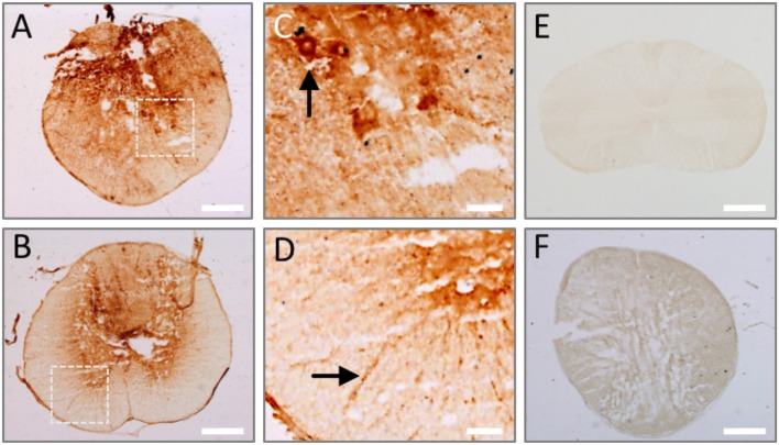
Figure 5.
Endogenous IgGs are detected in the spinal cord at 1 day after injury. Tissue sections from rat spinal cord at 1 day after injury were incubated with anti-rat IgG antibody and its binding was revealed by DAB-peroxidase reaction. Endogenous rat IgG is detected and depicts cellular profiles both in the lesion epicenter (A) and periphery (B). (C,D) Show magnified details of inserts in (A,B), where cell profiles in the ventral horn (arrow in C) and white matter (arrow in D) are clearly marked by endogenous IgG. (E) No endogenous IgG is observed in intact spinal cord. (F) IgG signal is species-specific, as shown by the absence of immune detection when using a secondary anti-rabbit IgG on injured spinal cord. Scale bar, 500 μm in (A,B,E,F); 100 μm in C and D.

