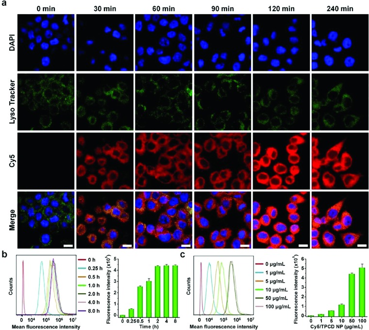
Figure 3.

Cellular uptake of Cy5‐labeled TPCD NP in RAW264.7 macrophages. a) Fluorescence images showing time‐dependent internalization of Cy5/TPCD NP at 2 µg mL−1 of Cy5 in RAW264.7 cells. For observation by confocal microscopy, nuclei were stained with DAPI (blue), while late endosomes and lysosomes were stained with LysoTracker (green). Scale bars, 10 µm. b) Typical flow cytometric curves (left) and quantitative analysis (right) of time‐dependent cellular uptake of Cy5/TPCD NP at 2 µg mL−1 of Cy5 in RAW264.7 cells. c) Flow cytometric profiles (left) and quantification results (right) indicating cellular uptake of Cy5/TPCD NP at various doses after 2 h of incubation in RAW264.7 cells. Data are mean ± SE (n = 3).
