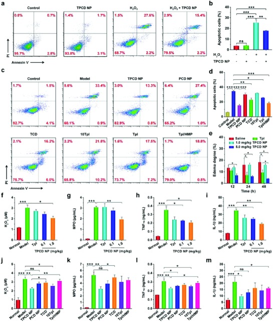
Figure 4.

In vitro antioxidative stress and in vivo anti‐inflammatory activities in murine models of acute inflammation. a,b) Flow cytometric profiles and quantitative data of apoptotic RAW264.7 cells after different treatments. The control, TPCD NP, and H2O2 groups were treated with fresh medium, 100 µg mL−1 of TPCD NP, and 200 × 10−6 m H2O2, respectively. The H2O2 + TPCD NP group was first incubated with 100 µg mL−1 of TPCD NP for 2 h, followed by culture with 200 × 10−6 m H2O2 for 24 h. c,d) Flow cytometric profiles and quantitative analysis of apoptotic macrophages treated with different formulations. The groups of TPCD NP, PCD NP, TCD, 10Tpl, Tpl, and Tpl/HMP were preincubated with the corresponding formulations for 2 h, and then stimulated with 200 × 10−6 m H2O2 for 24 h. The 10Tpl group was treated with Tpl at the tenfold dose of that contained in TPCD NP. e) In vivo efficacy of TPCD NP in rats with carrageen‐induced edema. At 0.5 h after challenge by i.d. injection of carrageen, a single dose of different formulations was locally administered. The dose of free Tpl was the same as that of TPCD NP at 5.0 mg kg−1. f–i) The levels of H2O2, MPO, TNF‐α, and IL‐1β in cell‐free peritoneal exudates collected from mice with zymosan‐induced peritonitis. At 1 h after zymosan induction by i.p. injection, different treatments were performed. TPCD NP at 1.0 mg kg−1 contained the same dose of Tpl as the free Tpl group. j–m) The expression of H2O2, MPO, TNF‐α, and IL‐1β in peritoneal exudates from peritonitis mice after treatment with different controls. Data are mean ± SE (b, d, n = 3; e, n = 5; f–m, n = 6). Statistical significance was assessed by one‐way ANOVA with post hoc LSD tests for data in (b, e–m). *P < 0.05, **P < 0.01, ***P < 0.001; ns, no significance. Due to heterogeneity of variance, the Kruskal–Wallis test was used for statistical analysis of data in (d). *P < 0.001, **P < 0.0001, ***P < 0.00001.
