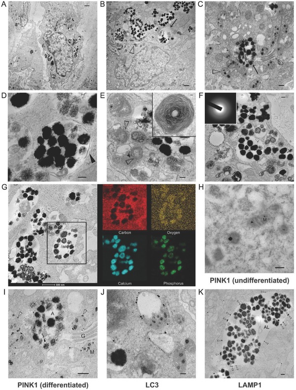
Figure 2.

TEM images illustrating the relationship between the electron‐dense granules within mitochondria and those within the cytosolic vesicles of hDPSCs. A) hDPSCs after exposure to ODM for 2 weeks (bar: 1 µm). B) High magnification of the intramitochondrial ACP granules (open arrowhead) and intravesicular granules (arrow) (bar: 50 nm). C) Coexistence of intramitochondrial ACP granules (open arrowhead), autolysosome (arrow), and Golgi apparatus (pointer) (bar: 200 nm). D) An autolysosome containing electron‐dense granules and other unidentifiable cytosolic components (bar: 200 nm). Partial degradation of the inner membrane derived from the original autophagosome could be seen (arrowhead). E) Multilamellar bodies identified (open arrowheads) adjacent to autolysosomes (bar: 200 nm). The inset shows a multilamellar body in higher magnification (bar: 50 nm). F) Granule‐containing autolysosomes in the vicinity of lysosomes (pointer) (bar: 200 nm). The electron‐dense granules were amorphous (inset). The granules were very close to the extracellular matrix. G) STEM‐EDX elemental mapping of the intravesicular electron‐dense granules revealed the presence of calcium, phosphorus, and oxygen, suggesting that those granules were ACP. H–K) Immunogold labeling of hDPSCs (bars: 200 nm for all images). H) In undifferentiated mitochondria (M) without granules, PINK1 was located within the interior of the mitochondria. I) In mitochondria destined for degradation, PINK1 was predominantly located (open arrowheads) along the periphery of the mitochondria (M). Some PINK1 could be identified around degraded organelles (solid arrowhead) within the autolysosome (A). G: Golgi body. J) LC3 was predominantly located along the membrane of mitochondria‐containing autophagosomes. K) LAMP1 was predominantly located along the membrane (open arrowheads) of the granule‐containing autolysosomes (AL).
