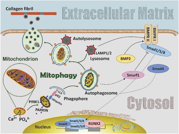
Figure 5.

Schematic of involvement of mitophagy in the transportation of amorphous calcium phosphate (ACP) precursors from the mitochondria to the extracellular matrix in stem cells with osteogenic potential. Calcium and phosphate ions are sequestered into the mitochondria matrices and accumulate as ACP granules. When ACP accumulation reaches a threshold, the mitochondria swell and lose membrane potential, resulting in PINK1 accumulation on their outer membrane. The latter recruits PARKIN from the cytosol to the damaged mitochondria. PARKIN ubiquitylates mitochondrial proteins and causes the ACP‐overloaded mitochondria to be engulfed by autophagosomes through p62 and LC3. The autophagosomes then fuse with lysosomes to become autolysosomes. Digestion of the dysfunctioned mitochondria by autolysosomal acidification releases the intramitochondrial ACP droplets, which coalesce into larger ACP granules within the autolysosomes. The coalesced ACP granules are subsequently transferred from the autolysosomes to the ECM via exocytosis. Infiltration of ACP into collagen fibrils results in intrafibrillar mineralization of the extracellular matrix. During this process, the BMP/Smad signaling pathway is activated to support the ACP transportation role of mitophagy in cell‐mediated osteogenesis. Abbreviations for pathway modification within the cytosol: solid black arrow: direct stimulatory modification; dashed black arrow: tentative stimulatory modification; dotted black arrow: joining of subunits and translocation; solid folding line with arrow: transcriptional stimulatory modification; solid line with right vertical bar: direct inhibitory modification. BMRR I and BMRR II: bone morphogenetic protein receptor I and II.
