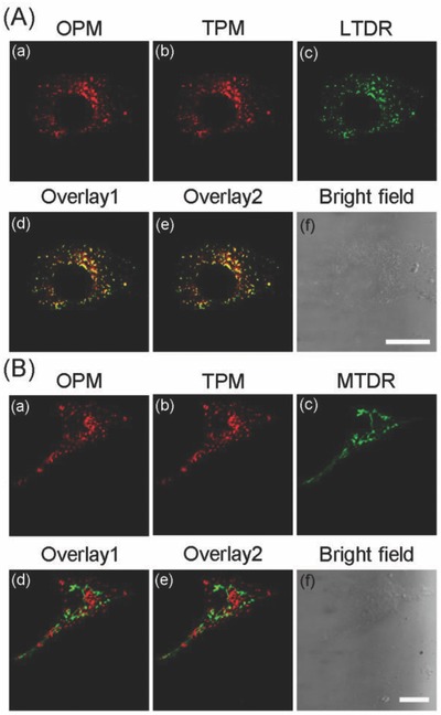
Figure 4.

U87 cells were colabeled with LysoIr@PDA‐CD‐RGD (1 × 10⁻⁶ m based on the concentration of LysoIr, 1 h) and A) LTDR (150 × 10−9 m , 0.5 h) or B) MTDR (50 × 10−9 m, 0.5 h). MTDR/LTDR: λex = 633 nm, λem = 665 ± 20 nm; OPM (one‐photon microscopy): λex = 405 nm, λem = 680 ± 20 nm; TPM (two‐photon microscopy): λex = 810 nm, λem = 680 ± 20 nm. (d) Overlay of (a) and (c). (e) Overlay of (b) and (c). Scale bars: 10 µm.
