Figure 5.

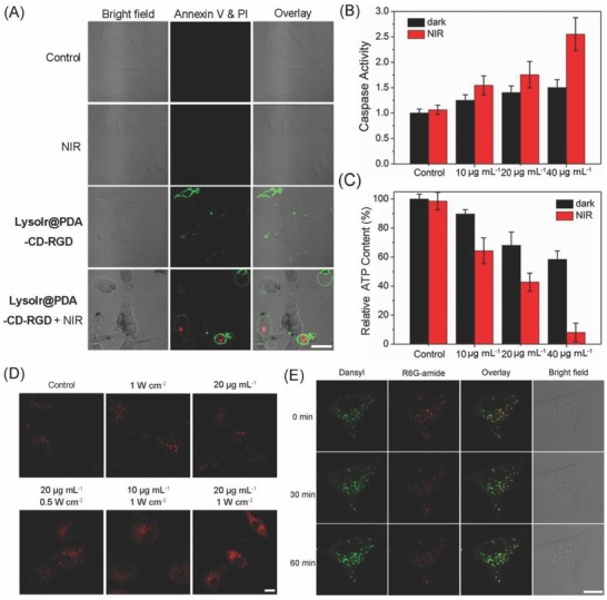
A) Representative confocal microscopic images of annexin V‐FITC/PI double stained U87 cells treated with LysoIr@PDA‐CD‐RGD (20 µg mL−1, 12 h) in the absence and presence of light. FITC: λex = 488 nm; λem = 530 ± 20 nm; PI: λex = 514 nm; λem = 620 ± 20 nm. B) Detection of caspase 3/7 activity in U87 cells treated with LysoIr@PDA‐CD‐RGD at the indicated concentrations in the absence or presence of light. C) Detection of cellular ATP content. D) Observation of cathepsin B release from lysosomes to cytosol in U87 cells treated with LysoIr@PDA‐CD‐RGD in the absence or presence of the 808 laser irradiation. λex = 543 nm; λem = 630 ± 20 nm. E) Visualization of lysosomal pH in U87 cells. The cells were treated with LysoIr@PDA‐CD‐RGD (20 µg mL−1, 4 h) and stained with Lyso‐DR (1 µg mL−1, 30 min). After irradiation, the cells were imaged with a confocal microscope at 0, 30 and 60 min. Dansyl: λex = 405 nm; λem = 440 ± 30 nm. R6G‐amide: λex = 543 nm; λem = 590 ± 30 nm. For the light‐treated samples in (A)‒(E), cells were irradiated with an 808 nm laser at a light dose of 1 W cm−2 for 5 min. Scale bar: 10 µm.
