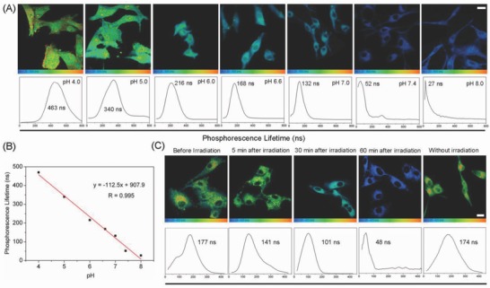
Figure 6.

A) TP‐PLIM images of U87 cells incubated with LysoIr (2 × 10⁻⁶ m, 1 h) at different pH values. λex = 810 nm; λem = 680 ± 20 nm. B) Linear fit between phosphorescence lifetimes of LysoIr and pH values in U87 cells. C) TP‐PLIM images of U87 cells incubated with LysoIr@PDA‐CD‐RGD. The cells were treated with LysoIr@PDA‐CD‐RGD (20 µg mL−1, 2 h) and then irradiated with an 808 laser (1 W cm−2, 5 min). Scale bar: 10 µm.
