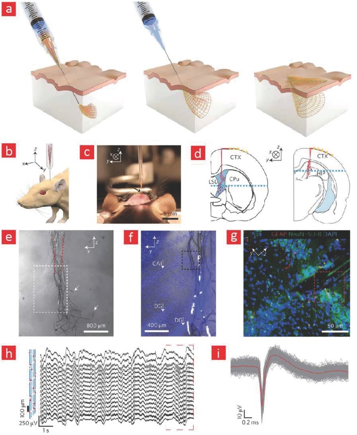
Figure 15.

Syringe‐injectable electronics for neural recording. a) Depictions of syringe‐injectable electronics. b) Schematics showing the injection of the electronics into the brain of mice. c) Photographs depicting the injection process into a three‐month‐old mouse brain. d,e) Schematics showing the areas of the mice brain, wherein the mesh electronics was injected. f) Bright‐field microscopy imaging of the brain region into which the mesh electronics was injected five weeks after injection. g) Bright‐field and epi‐fluorescence images corresponding to the region indicated by a white box in (f). h) Fluorescence image corresponding to the region indicated by a blue box in (f). i,j) Electrical recordings from the mouse brain using the injected mesh electronics. Adapted with permission.409 Copyright 2015, Macmillan Publishers Ltd.
