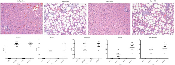
Fig. 1.
Liver histopathology of control and MCD-diet rats. H&E-stained liver sections of control rats and rats fed 8 weeks of MCD diet, WT, and Bcrp KOs. Hallmark characteristics of NASH were found in MCD rats, whereas control group animals showed no signs of NASH pathology. Original magnification, 40×. Two-way analysis of variance, *P ≤ 0.05; **P ≤ 0.01; ***P ≤ 0.001, n = 3, n = 4 (KO-MCD).

