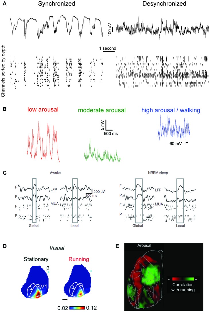
Figure 1.
An updated definition of brain states. (A) Brain states have traditionally been distinguished based on the characteristic of population-level oscillatory dynamics present within a behaviorally-defined homogeneous period. Left, top: local field potential (LFP) trace present in mouse visual cortex during isoflurane anesthesia. LFP activity is characterized by oscillatory dynamics with a strong power in slow frequencies (0.5–4 Hz). Left, bottom: at the neuronal level, isoflurane anesthesia determines the alternation of periods of spiking and silence which are synchronous throughout cortical areas, and in phase with the co-occurring LFP oscillations. Right: same as left panel, for activity typical of wakefulness. Note the disappearance of slow frequency oscillations at the LFP level and the overall loss of synchrony for neuronal activity. (B) Recent studies have shown that, within wakefulness, much more variability is present than previously thought. In periods characterized by a low arousal level (left) neuronal activity (here shown in terms of intracellular membrane potential traces) displays patterns indistinguishable from those present during Non-REM sleep or anesthesia. As the arousal level increases, or if a period of activity—locomotion—occurs (center, right) activity becomes more and more desynchronized. Wakefulness can thus be subdivided in several micro-states, with markedly different properties. Adapted from McGinley et al. (2015b), permission to reproduce has been obtained from the publisher. (C) During Non-REM sleep and forms of non-dissociative (e.g., isoflurane) anesthesia UP and DOWN states normally occurs globally, i.e., involving the whole thalamocortical system. However, UP/DOWN states can also be local events, involving one a set of cortical regions. This mostly occurs during wakefulness when homeostatic sleep pressure is high (left), and during Non-REM sleep when, conversely, sleep pressure is slow. Each plot shows an example of global or local DOWN state occurring during wakefulness (left) or sleep (right), and measured in terms of both LFP recordings or neuronal multi-unit activity (MUA). F: frontal derivation. P: parietal derivation. Adapted with permission from the authors from Vyazovskiy et al. (2011). (D) Sensory processing within wakefulness varies across distinct micro-states. Visual evoked potentials (here shown as measure by voltage-sensitive fluorescent proteins) are generally weaker when a mouse is running compared to when it is stationary. The same is observed for audition and somatosensation. B: Bregma. Adapted under Creative Commons Attributions CC BY 4.0 license from Shimaoka et al. (2018). (E) Arousal differentially affects cortical areas in the mouse. Green indicates cortical areas where the signal measured via voltage-sensitive fluorescent protein is positively correlated with arousal (locomotion). Red shows areas where a negative correlation is present. Adapted under Creative Commons Attributions CC BY 4.0 license from Shimaoka et al. (2018).

