FIG 4.

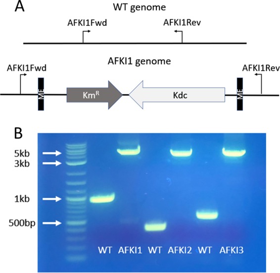
The transposon integration sites were confirmed by amplification of the region of the genome flanking the insertion sites. Panel A shows the genomic PCR amplification scheme for the AFKI1 strain. The primers flanking the insertion site were designed from the published genome sequence for ATCC 23270. In the wild-type genome, the PCR amplicon is of a known size. In the transposon-integrated strains, the same PCR primers were used to amplify the transposon region as well, resulting in a 3.5-kb increase in amplicon size. Panel B shows the results of the method applied to the three transposon locations identified in the mutant strains and visualized on an agarose gel, confirming the successful identification of integration sites.
